

When To Worry About MRI Results
When To Worry About MRI Results
Reviewed by Otto R. Garcia MD Internist at Hermosa Medical and Diagnostic Center
Last updated: September 2026
Key takeaways
- Most abnormal MRI findings are age related wear rather than disease.
- Read the impression section first. Findings not repeated there were judged unimportant.
- Radiologists must phone critical findings to the ordering doctor usually within the hour.
- Routine reports take 24 to 72 hours. Silence almost always means nothing urgent was found.
- 9 specific phrases warrant a call within 48 hours rather than waiting for a routine appointment.
Most abnormal MRI findings are not dangerous. Words like degenerative change disc bulge signal alteration and nonspecific describe normal wear present in a large share of healthy people with no symptoms at all.
Worry is warranted for a small set of findings. A mass or lesion described as enhancing infiltrative or ill defined. Anything called suspicious for malignancy. Cord compression. Acute infarct. Fracture. Abscess. These appear in the impression section rather than buried in the body of the report.
If something on your MRI scan is genuinely urgent you will usually hear within hours and not days. Radiologists are required to call the ordering physician directly for critical findings. Silence after a scan almost always means nothing urgent was found.
Report language decoded
|
Phrase in the report |
What it means |
Concern level |
What usually happens next |
|
Unremarkable |
Normal |
None |
Nothing |
|
No acute abnormality |
Nothing new or urgent |
None |
Nothing |
|
Stable compared with prior study |
No change since the earlier scan |
Very low |
Routine follow up at most |
|
Degenerative change |
Age related wear of discs or joints |
Very low |
Treat symptoms not the image |
|
Nonspecific |
Appearance does not point to one cause |
Low |
Watch or correlate with symptoms |
|
Likely benign or probably benign |
Recognized low risk category |
Low |
Interval follow up scan |
|
Incidental finding |
Found by accident and unrelated to the scan reason |
Low |
Usually documentation only |
|
Enhancing or avidly enhancing |
Tissue takes up contrast. Increased blood supply |
Moderate |
Further imaging or specialist referral |
|
Suspicious for or concerning for |
A specific worry is being named |
High |
Prompt follow up. Sometimes biopsy |
|
Cord compression |
Pressure on the spinal cord |
Urgent |
Same day surgical assessment |
|
Acute infarct |
New tissue death from blocked blood supply |
Urgent |
Immediate treatment |
Read a phrase in your report you do not recognize? Ten minutes with the doctor who ordered the scan usually settles it. Call 773 772 8876 or book an appointment online.
How to actually read an MRI report
Every radiology report has the same four part structure. Clinical history. Technique. Findings. Impression. Reading them in the right order prevents most unnecessary panic because the findings section is an exhaustive inventory and the impression is the judgement.
Clinical history. One line describing why the scan was ordered. This shapes what the radiologist looked for.
Technique. Which sequences were run and whether contrast was used. Mostly technical.
Findings. A long descriptive inventory of everything visible. This section describes and does not interpret. It lists incidental things a radiologist is obliged to mention even when they mean nothing at all.
Impression. The radiologist conclusion. This is the section that matters. It states what is significant what is probably not and what should happen next.
Read the impression first. If a finding appears in the findings section and not in the impression the radiologist decided it was not clinically important. That single habit resolves a great deal of anxiety. Our article on what impression means on an MRI report covers that section in depth.
Language that should reassure you
Eight phrases appear constantly in MRI reports and signal a low concern finding. Degenerative change. Unremarkable. No acute abnormality. Mild disc bulge. Nonspecific white matter changes. Likely benign. Incidental finding. Stable compared with prior study.
Degenerative change. Age related wear. Nearly universal after forty. Not a disease.
Mild disc bulge or disc desiccation. Common in people with no back pain at all. Imaging studies of asymptomatic adults find disc bulges in a large share of participants in their forties and fifties.
Nonspecific white matter changes. Small bright spots on brain MRI. Extremely common with age migraine and blood pressure history and usually not significant alone.
Likely benign or probably benign. A recognized radiology category meaning the chance of anything serious is very low. Standard management is a follow up scan rather than a biopsy.
Incidental finding or incidentaloma. Something found by accident that had nothing to do with why you were scanned. Most need nothing beyond documentation.
Stable compared with prior study. One of the most reassuring phrases available. Stability over time is strong evidence against an aggressive process.
Language that warrants a call within 48 hours
Nine terms mean the report needs discussion soon rather than at your next routine visit. None of them automatically mean bad news. They mean a conversation is due.
Mass or lesion. Neutral descriptive words covering both benign and malignant findings. Context in the impression decides.
Enhancing or avidly enhancing. The tissue takes up contrast indicating increased blood supply. Tumours inflammation and infection all enhance.
Suspicious for or concerning for. The radiologist is naming a specific worry. Follow up is expected.
Ill defined or infiltrative margins. Aggressive processes tend to have unclear borders. Benign ones tend to have sharp borders.
Cord compression or canal stenosis with cord signal change. Pressure on the spinal cord. This can need urgent surgical assessment particularly with weakness numbness or bowel and bladder changes.
Acute infarct. New tissue death from blocked blood supply. Urgent.
Restricted diffusion. Often indicates acute stroke abscess or a densely cellular tumour. Always read in context.
Fracture or marrow signal change. New bone injury.
Recommend tissue sampling or biopsy. A direct recommendation for the next step.
How fast results arrive and what silence means
A typical outpatient MRI is read within 24 to 72 hours. The report reaches the ordering physician who reviews it and contacts you. If something critical appears the radiologist does not wait for that cycle. Professional standards require direct communication with the ordering clinician for critical results often within an hour.
This is why the common fear that bad news would be hidden until an appointment is largely unfounded.
Patient portals complicate the picture. Under current United States information blocking rules results are frequently released to the portal immediately on finalization. Many patients read the report before their doctor has called which produces a predictable pattern where someone reads the word lesion at eleven at night and spends a sleepless night over a finding their physician would have described as unimportant in ten seconds.
Two practical rules help. Read the impression and not the findings. Then wait for the interpretation rather than searching individual phrases because search engines return worst case explanations for neutral descriptive terms.
If four business days pass with no contact call the ordering office and ask. Reports do occasionally get missed in a workflow and a phone call resolves it.
Why an MRI finding often does not explain your pain
Imaging finds structural changes and structural changes correlate loosely with symptoms. Studies scanning people with no back pain consistently find disc degeneration bulges and facet arthritis at high rates that rise steadily with age. A finding on your scan may be a coincidence rather than the cause.
The implication matters. Treating an image instead of a patient leads to procedures that do not help and occasionally to surgery for a finding that was never producing symptoms.
The reverse also happens. Severe pain with a clean MRI is real. Nerve irritation without structural compression muscular pain and central sensitization all produce genuine pain that MRI cannot visualize. A normal scan is not a statement that the pain is imagined.
Your physician weighs the image against your symptoms your examination and your history. That reconciliation is the actual diagnosis. The scan is one input into it and not a verdict on its own. Our internal medicine team does that reconciliation in the same building where the scan was taken.
Chicago winter back pain and what scans show
Back pain complaints rise across Chicago every winter and the causes are mostly mechanical rather than structural. Snow shovelling produces a recognizable cluster of acute back injuries every season. Ice related falls produce another. Reduced outdoor activity across four or five months deconditions the muscles that support the spine.
The practical consequence for imaging is worth understanding. Someone scanned in February after a shovelling injury will very often show degenerative changes that were present long before the injury and will be present long after it resolves.
Seeing those words on a report after an acute injury leads people to conclude the injury caused permanent damage. Usually it did not. The wear was already there and the scan simply documented it.
This pattern shows up constantly across Hermosa Belmont Cragin and Logan Square between January and March. The useful question to ask your doctor is whether the finding is new or whether it looks like background wear.
A typical case we see
A man in his sixties from Austin read his own lumbar spine report on a patient portal at midnight. It described multilevel degenerative disc disease facet arthropathy and a disc protrusion. He searched each phrase and arrived at his appointment convinced he needed surgery.
The impression also contained the words no nerve root compression. Those four words were the whole answer and they had not registered among the alarming vocabulary above them.
His pain was mechanical and it responded to physical therapy across about eight weeks. The scan findings were background wear typical for his age. The case is common enough to be worth stating plainly. Reading the findings section without the impression produces unnecessary fear more often than it produces useful understanding.
Illustrative example. Composite of common presentations. Not a specific individual and not a testimonial. replace with a real anonymized case from clinic records.
Call 773 772 8876 or book an appointment online
See our Radiology and imaging services page for what the visit covers
Hablamos español. Llame al 773 772 8876 para programar su cita.
What to ask at the follow up appointment
Bring seven questions written down because appointments move quickly and people forget what they meant to ask.
What is the single most important finding on this scan.
Does this finding explain the symptoms I came in with or is it unrelated.
Is this finding new or was it present on an earlier study.
What is the realistic list of possible causes and which is most likely.
What happens next and what is the timeline.
If we watch this instead of treating it what specifically are we watching for.
What symptoms should make me call before the next appointment.
Ask for a copy of the images on disc or through the portal as well as the report. If you seek a second opinion the images matter more than the text because another radiologist needs to look at them rather than read someone else's description.
When an MRI finding is genuinely urgent
Seven symptoms alongside a recent scan mean immediate care rather than a scheduled appointment. New weakness in an arm or leg. Loss of bladder or bowel control. Numbness in the saddle region between the legs. Sudden severe headache unlike any previous headache. New confusion or difficulty speaking. Fever with back pain and a spinal finding on imaging. Progressive difficulty walking.
These combined with findings such as cord compression epidural collection or acute infarct are emergencies regardless of what any appointment calendar says.
Do not wait for a callback in these situations. Go to an emergency department and bring the report and the disc if you have them.
The reason this list matters is that patients frequently receive a report describing cord compression read it as concerning and book a routine appointment three weeks out. Cord compression with new weakness is a same day problem.
Getting a second reading
Second opinions on imaging are reasonable and common particularly for cancer staging complex spine findings and any recommendation for major surgery.
Bring the actual images and not only the report. A second radiologist reading the same images sometimes reaches a different conclusion which is exactly why the disc matters more than the text.
Ask whether comparison with any prior imaging was performed. Comparison changes interpretation more than almost anything else and reports frequently state no prior studies available simply because the images were never requested from the other facility.
Retrieving prior images can convert an uncertain finding into a stable one and occasionally removes the need for a follow up scan entirely. Bring a list of where you have been imaged before.
What happens at your appointment
Expect 30 to 45 minutes for the scan itself and a separate shorter visit to review results.
Booking and prior authorization. Most insurers require prior authorization for MRI and it is the commonest cause of delay. Start it as soon as the scan is ordered.
Check in. Photo identification insurance card and your implant list. Arrive fifteen minutes early for screening paperwork. Staff speak English Spanish and Arabic.
Metal screening. Pacemakers defibrillators cochlear implants aneurysm clips insulin pumps and metal fragments in the eye all require review. Bring implant cards. Many modern devices are MRI conditional meaning safe under specific protocols.
Changing. Jewellery watches and hair accessories removed. Clothing with metal including underwire and zippers usually means changing into a gown.
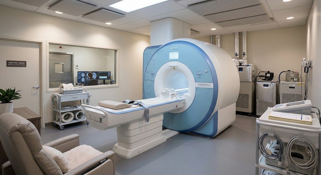
The scan. 20 to 45 minutes depending on the region. Ear protection provided. Our open MRI suite means you are not enclosed in a tube which matters for anyone with claustrophobia.
Contrast if ordered. Given intravenously. Kidney function is checked first where indicated.
Report. Read within 24 to 72 hours. Critical findings are phoned to the ordering physician far sooner.
Results review. Booked with the physician who ordered the scan. Bring your questions written down.
What Medicaid and Medicare cover for MRI scans and imaging in Chicago
Coverage depends on whether a service is medically necessary or cosmetic and it varies by plan. The summary below reflects how Illinois Medicaid and Medicare generally treat these services. Confirm your own plan before the visit because benefits differ between managed care plans.
Generally covered
- MRI ordered by a treating physician for a documented clinical indication
- Contrast agent where clinically indicated
- Radiologist interpretation and report
- Follow up imaging recommended by the radiologist
- Medicare Part B and Illinois Medicaid both cover medically necessary MRI
Generally not covered
- Whole body MRI performed as screening without a clinical indication
- Imaging ordered without a physician order in most plans
- Repeat imaging at a second facility for a second opinion in many plans
Worth bundling into the same visit
- Office visit with the ordering physician at the same trip
- Blood work including kidney function before contrast
- X ray or ultrasound where both are indicated
- Specialist referral arranged at the same visit
We accept Medicaid and Medicare. Call 773 772 8876 to confirm your plan before the visit. Coverage varies by plan so call to confirm rather than assuming.
Frequently asked questions
If my MRI results are bad do they tell me right away?
Yes in the great majority of cases. Radiologists are required to communicate critical findings directly to the ordering physician usually within an hour. Routine results take 24 to 72 hours.
What does nonspecific mean on an MRI report?
It means the appearance does not point clearly to one cause. It is a hedge rather than a warning and such findings are usually followed rather than treated.
Does the word lesion on an MRI mean cancer?
No. Lesion means an area of abnormal tissue. Cysts scars inflammation benign tumours and infections are all lesions. The impression section tells you which concern applies.
Which MRI findings are urgent?
Cord compression acute infarct abscess fracture and anything described as suspicious for malignancy. New weakness loss of bladder or bowel control or saddle numbness require immediate care.
Why does my MRI finding not explain my pain?
Disc bulges and degenerative change appear commonly in people with no symptoms at all. Imaging correlates loosely with pain so your physician weighs the scan against your examination and history.
How long does an MRI report take?
Most outpatient MRI studies are read within 24 to 72 hours. Critical findings are phoned to the ordering physician far sooner.
Does Medicaid cover MRI in Illinois?
Yes for medically necessary imaging ordered by a treating physician with documented indication. Prior authorization is commonly required. Call 773 227 9777 for our imaging centre or 773 772 8876 for the main clinic.
Should I get a second opinion on my MRI?
Reasonable for cancer staging complex spine disease or before major surgery. Bring the images themselves and not just the report because another radiologist needs to look at them.
About the reviewer
Otto R. Garcia MD | Internist | Hermosa Medical and Diagnostic Center
Dr Garcia is a board certified internal medicine specialist at Hermosa Medical Center. He cares for adults across the full range of chronic and acute conditions with a focus on long term disease management. He sees patients in English and Spanish.
Imaging at Hermosa Medical and Diagnostic Center is performed at the on site radiology centre and scans are interpreted by a reporting radiologist. This article was medically reviewed by a board certified physician on staff who orders and acts on these scans. A radiologist byline will replace this line once the clinic supplies the reporting radiologist name and credential.
Next step
If you are holding a report you do not understand book a short review visit rather than searching individual phrases. The impression section usually contains the answer and it takes ten minutes to walk through.
Go to Radiology and imaging services to see what we offer and how to book.
Visit Hermosa Medical and Diagnostic Center
Hermosa Medical and Diagnostic Center has served the Chicago West Side for 33 years. Primary care specialty care laboratory services imaging and an on site pharmacy sit in one building so most visits finish in one trip.
Address 2004 N Pulaski Rd Chicago IL 60639
Phone 773 772 8876
Advantage MRI 773 227 9777
Hours Call 773 772 8876 to confirm current hours
Walk ins Welcome. No appointment needed.
Insurance Medicaid Medicare and most major plans accepted
Languages English Spanish and Arabic
We serve Hermosa Belmont Cragin Humboldt Park Logan Square and Austin.
*Hablamos español. Llame al 773 772 8876 para programar su cita. Se habla español.*
Getting here
We are at 2004 N Pulaski Rd in the Hermosa neighborhood just north of Armitage Ave. Street parking is available on Pulaski Rd and on the surrounding side streets. The CTA 53 Pulaski bus stops directly outside and the 73 Armitage bus runs east from the corner toward the Blue Line.
Embedded Google Map of 2004 N Pulaski Rd Chicago IL 60639. Open in Google Maps (the map embed code is in the markdown version of this file).